Physiology
Right now, as you read this sentence, your digestive system is breaking down your last meal into molecules small enough to enter individual cells. Your heart is beating approximately 70 times per minute. Your lungs are exchanging gases 15 times every minute. Your nervous system is processing millions of signals simultaneously.
All of this is happening without a single conscious thought from you.
Physiology is the branch of biology that studies exactly how all of this works. Not what the parts are named or where they sit in the body, but what they actually do, how they do it, and how they coordinate with each other. It is the science of life in action, and it is what turns anatomy from a map into a story.
Physiology of the Digestive System
The digestive system converts the complex molecules in food into small, soluble molecules that can be absorbed into the bloodstream and distributed to cells throughout the body.
Mechanical and Chemical Digestion
Digestion involves two complementary processes working together.
Mechanical digestion:
- Physically breaks food into smaller pieces
- Increases surface area for chemical digestion
- Occurs through chewing in the mouth and churning in the stomach
Chemical digestion:
- Uses enzymes to break chemical bonds within large food molecules
- Converts large molecules into their smaller building blocks
- Occurs throughout the digestive tract
The Mouth and Esophagus
- Teeth mechanically break food into smaller pieces
- Salivary glands secrete saliva containing salivary amylase
- Salivary amylase begins hydrolysis of starch into maltose
- Food is shaped into a rounded mass called a bolus and swallowed
- The bolus travels down the esophagus through peristalsis
- Peristalsis consists of coordinated waves of contraction and relaxation in smooth muscle
- Pushes food toward the stomach regardless of body position
The Stomach
- Gastric glands secrete hydrochloric acid, creating a pH of 1.5 to 3.5
- Acid kills most pathogens in food
- Acid denatures proteins, making them more accessible to enzymes
- Acid activates pepsin from its inactive precursor, pepsinogen
- Pepsin begins hydrolysis of proteins into shorter polypeptide chains
- Muscular walls churn food into a semi-liquid paste called chyme
- Chyme is gradually released into the small intestine through the pyloric sphincter
The Small Intestine
The small intestine is where the majority of chemical digestion and all absorption takes place.
Secretions entering the duodenum:
From the pancreas:
- Pancreatic amylase: completes starch digestion to maltose
- Lipase: hydrolyzes triglyceride fats into fatty acids and glycerol
- Proteases, including trypsin, complete protein digestion to amino acids
From the liver via the gallbladder:
- Bile emulsifies fat into tiny droplets
- Increases surface area for lipase to work on
- Bile is not an enzyme and does not chemically digest fat
Adaptations for absorption:
- Wall folded into thousands of finger-like villi
- Each villus cell is covered in microvilli, forming the brush border
- Total absorptive surface area is approximately 200 square meters in a tube only 6 meters long
How products are absorbed:
- Glucose and amino acids: absorbed by transport proteins into capillaries within villi, transported to the liver via the hepatic portal vein
- Fatty acids and glycerol: reassembled into triglycerides, packaged into chylomicrons, and enter lymphatic capillaries within each villus
The Large Intestine
- Water is reabsorbed from the remaining indigestible material
- Bacteria ferment some fiber, producing vitamins B and K
- The remaining material is compacted and stored as feces before elimination
Physiology of the Circulatory System
The circulatory system transports materials around the body, delivering oxygen and nutrients to every cell and collecting carbon dioxide and metabolic waste for removal.
The Heart
The heart is a muscular pump divided into four chambers.
Chambers and their roles:
- Right atrium: receives deoxygenated blood from the body
- Right ventricle: pumps deoxygenated blood to the lungs
- Left atrium: receives oxygenated blood from the lungs
- Left ventricle: pumps oxygenated blood to the rest of the body
This arrangement creates double circulation, ensuring oxygenated and deoxygenated blood remain completely separated and that blood reaches body tissues at high pressure.
The Cardiac Cycle
Diastole:
- Heart relaxes
- Chambers filled with blood
- Atrioventricular valves open
Systole:
- Ventricles contract powerfully
- Blood pushed out through the arterial exits
- Valves prevent backflow
Heartbeat initiation:
- The sinoatrial node (SA node) in the right atrium generates rhythmic electrical impulses
- Impulses spread across both atria, causing simultaneous contraction
- Signal reaches the atrioventricular node (AV node), which delays it briefly
- Impulse travels through conducting fibers to the ventricles
- Ventricles contract from bottom up, squeezing blood out
Blood Vessels
| Vessel |
Direction |
Wall |
Special Features |
| Arteries | Away from heart | Thick, muscular, elastic | Withstand high pressure |
| Veins | Toward heart | Thinner | Valves prevent backflow |
| Capillaries | Through tissues | One cell thick | Site of all exchange |
Blood Composition
- Plasma: Liquid matrix carrying dissolved nutrients, hormones, and waste products; transports carbon dioxide dissolved and as bicarbonate ions
- Red blood cells: Biconcave disc shape maximizes surface area for gas exchange; no nucleus in mature form allowing maximum hemoglobin content; hemoglobin binds oxygen in lungs and releases it in tissues
- White blood cells: Components of the immune system; various types with different roles in immune defense
- Platelets: Small cell fragments that initiate blood clotting when vessel walls are damaged
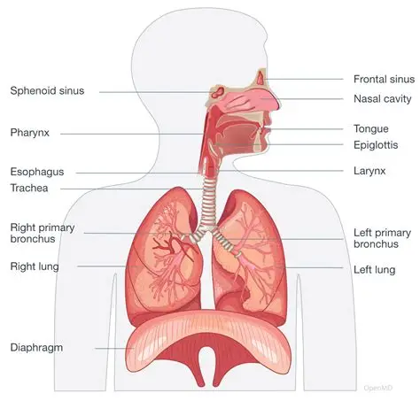
Physiology of the Respiratory System
The respiratory system brings oxygen into the body and removes the carbon dioxide produced by cellular respiration in tissues.
Mechanics of Breathing
Inhalation:
- The diaphragm contracts and flattens
- External intercostal muscles contract, pulling the ribcage upward and outward
- The volume of the thorax increases
- Pressure inside the lungs falls below atmospheric pressure
- Air flows in from outside
Exhalation at rest:
- The diaphragm and intercostal muscles relax
- The ribcage falls inward and downward
- Thoracic volume decreases
- Pressure rises above atmospheric
- Air flows out
Forced exhalation during exercise: Internal intercostal muscles and abdominal muscles contract actively, further reducing thoracic volume for more complete expulsion of air.
Gas Exchange in the Alveoli
Gas exchange occurs in the alveoli, tiny air sacs deep within the lungs.
Structural adaptations for efficient gas exchange:
- Walls just one cell layer thick, minimizing diffusion distance
- Approximately 500 million alveoli providing surface area of 70 to 100 square meters
- Dense surrounding capillary network maintaining short diffusion path
- Steep concentration gradients are maintained by continuous blood flow and breathing
- Moist inner surfaces allow gases to dissolve before diffusing across epithelium
Gas movement:
- Oxygen diffuses from high concentration in alveolar air into lower concentration in blood, binds to hemoglobin, forming oxyhemoglobin
- Carbon dioxide diffuses from higher concentration in the blood into lower concentration in the alveolar air and is exhaled
Transport of carbon dioxide in blood:
- Dissolved in plasma
- Bound to hemoglobin
- As bicarbonate ions (approximately 70 percent of total)
Physiology of the Nervous System
The nervous system detects stimuli from the environment, processes information, and coordinates appropriate responses.
Neurons and Action Potentials
Neuron structure:
- Cell body: contains nucleus and metabolic machinery
- Dendrites: short branched extensions that receive incoming signals
- Axon: single long extension that conducts the electrical signal away from cell body
How an action potential works:
- At rest: Inside a neuron is negatively charged relative to the outside (resting potential)
- When a stimulus arrives: Sodium ion channels open; sodium ions rush in down their concentration gradient; inside briefly becomes positively charged (depolarization); this triggers the same process in the adjacent membrane region
- An action potential propagates along the axon
- Restoration: Potassium ions flow out to restore the resting potential
Myelin sheath:
- A fatty insulating sheath wrapped around many axons
- Forces the action potential to jump between the nodes of Ranvier
- Dramatically increases the speed of signal transmission
- Myelinated neurons conduct at up to 120 meters per second
Synaptic Transmission
Neurons communicate with each other at junctions called synapses.
Process:
- The action potential reaches the presynaptic terminal
- Triggers the release of neurotransmitters from vesicles into the synaptic cleft
- Neurotransmitters diffuse across the gap
- Bind to receptor proteins on the postsynaptic membrane
- Trigger a new action potential or inhibit one
- Neurotransmitters are rapidly broken down or reabsorbed
Reflex Arcs
A reflex is a rapid, automatic, involuntary response to a stimulus that does not require conscious processing by the brain.
Pathway of a reflex arc:
- The receptor detects a stimulus
- A sensory neuron carries a signal to the spinal cord
- A relay neuron in the spinal cord connects directly to a motor neuron
- A motor neuron activates an effector muscle or gland
- The brain receives information about the reflex after it has already occurred
The brain is bypassed in the initial response, which is why reflexes are faster than voluntary reactions.
Homeostasis: The Integration of All Physiology
Homeostasis is the maintenance of a stable internal environment despite continuous changes in external conditions and internal activity.
Temperature Regulation
Set point: Approximately 37 degrees Celsius
- When temperature rises: Thermoreceptors in the hypothalamus detect a change; blood vessels in the skin dilate to increase heat loss; sweat glands are activated to cool the skin through evaporative cooling
- When temperature falls: Blood vessels in the skin constrict to reduce heat loss; skeletal muscles shiver to generate heat through rapid contraction
Blood Glucose Regulation
- After eating (blood glucose rises): Beta cells in the pancreas detect rising glucose; insulin is secreted into the blood; liver and muscle cells convert glucose to glycogen for storage; other cells increase glucose uptake; blood glucose returns to the normal range
- Between meals (blood glucose falls): Alpha cells in the pancreas detect falling glucose; glucagon is secreted into the blood; the liver breaks down glycogen and releases glucose into the blood; blood glucose returns to the normal range
Negative Feedback
Both temperature regulation and blood glucose regulation operate through negative feedback.
The response to a deviation from the set point produces an effect that opposes the deviation and returns the variable toward its normal value.
In type 1 diabetes, Beta cells are destroyed by the immune system. No insulin is produced. Blood glucose rises to dangerously high levels without insulin injections.
In type 2 diabetes, Body cells become resistant to insulin. Glucose cannot enter cells efficiently even when insulin is present.
Negative feedback is the fundamental control principle underlying virtually all homeostatic mechanisms in the body. Understanding it explains how a living organism maintains internal stability in a world that is constantly changing around it.
Why Physiology Is the Heart of Biology
Physiology connects structure to function; it is not enough to know what the heart looks like if you don't understand how it pumps. It is not enough to know the alveoli are thin-walled without understanding why that thinness matters for diffusion. Every structural feature of every organ makes sense only in the context of its physiological function.
Physiology is also the foundation of medicine. Understanding how healthy systems function is essential for recognizing, diagnosing, and treating dysfunction — whether that dysfunction is a blocked artery, a diabetic pancreas, or a nerve injury. The living, working human body is physiology in constant action.