The Nervous System
Reach out and touch a hot surface. Before you have even consciously decided to move, your hand has already pulled away. A signal traveled from your fingertip to your spinal cord and back to your muscles in a fraction of a second, faster than your brain could process the event.
Now consider that as you read this page, billions of neurons in your brain are firing in coordinated patterns, encoding meaning, accessing memories, planning responses, and maintaining your awareness of everything around you.
The nervous system is the most complex biological structure known. It allows organisms to detect changes in their environment with extraordinary precision, process information at remarkable speed, and coordinate responses involving hundreds of different muscles and organs working in perfect synchrony.
Organization of the Nervous System
The human nervous system is organized into two main divisions.
Central Nervous System (CNS)
The central nervous system consists of the brain and spinal cord. It is the processing center of the nervous system, receiving sensory information, integrating it, and sending out instructions.
The brain and spinal cord are protected by:
- The skull and vertebral column (bony protection)
- Three layers of protective membranes called meninges
- Cerebrospinal fluid, which cushions against mechanical shock and provides nutrients
Peripheral Nervous System (PNS)
The peripheral nervous system consists of all the nerves outside the brain and spinal cord. It connects the CNS to every part of the body.
Somatic nervous system:
- Controls voluntary movements of skeletal muscles
- Carries sensory information from skin, muscles, and joints to the CNS
- Under conscious control
Autonomic nervous system:
- Controls involuntary functions of internal organs, glands, and smooth muscle
- Operates largely below the level of conscious awareness
The autonomic system is further divided into:
- Sympathetic division: Prepares the body for activity. Increases heart rate, dilates airways, redirects blood to muscles, and releases adrenaline. Dominant during stress and exercise (fight or flight).
- Parasympathetic division: Promotes rest and digestion. Slows heart rate, stimulates digestive activity, and conserves energy. Dominant during rest (rest and digest).
These two divisions work antagonistically, continuously adjusting organ function to match the body's current needs.
Neurons
Neurons are the specialized cells of the nervous system that generate and transmit electrical signals called action potentials.
There are approximately 86 billion neurons in the human brain alone.
Types of Neurons
- Sensory neurons (afferent): Carry signals from sensory receptors toward the CNS. Have long dendrites extending to peripheral receptors. The cell body is located in the ganglia outside the CNS.
- Motor neurons (efferent): Carry signals from the CNS to effectors (muscles and glands). Have long axons extending to muscles and glands. The cell body is located within the CNS.
- Relay neurons (interneurons): Located entirely within the CNS. Connect sensory and motor neurons. Form the complex processing networks of the brain and spinal cord. Most neurons in the brain are relay neurons.
Structure of a Motor Neuron
- Cell body (soma): Contains the nucleus and most metabolic machinery. Receives signals from dendrites.
- Dendrites: Multiple short, branched extensions that receive incoming signals from other neurons or sensory receptors. A single neuron may have thousands of dendrites.
- Axon: A single long extension that carries the action potential away from the cell body toward the next neuron or effector. Can be up to a meter long in humans.
- Myelin sheath: A fatty insulating layer wrapped around many axons by Schwann cells. Speeds up signal transmission dramatically by forcing the action potential to jump between gaps called nodes of Ranvier (saltatory conduction). Myelinated axons conduct at up to 120 m/s. Unmyelinated axons conduct at only 0.5 to 2 m/s.
- Axon terminals (synaptic knobs): The branched endings of the axon where neurotransmitters are stored and released.
The Action Potential
An action potential is the electrical signal that travels along a neuron. It is an all-or-nothing event: either a full action potential is generated or none at all.
Resting Potential
When a neuron is not transmitting a signal, the inside of the cell membrane is negatively charged relative to the outside. This is the resting potential, approximately -70 mV.
This charge difference is maintained by:
- The sodium-potassium pump actively moves 3 Na⁺ out and 2 K⁺ in per ATP molecule
- The membrane is more permeable to K⁺ than Na⁺ at rest, allowing K⁺ to leak out
Depolarization
When a stimulus of sufficient strength arrives at the neuron:
- Voltage-gated sodium channels open
- Na⁺ ions rush into the cell down their concentration and electrical gradients
- The inside of the membrane becomes positively charged (depolarization)
- The membrane potential rises to approximately +40 mV
Repolarization
- Sodium channels close
- Voltage-gated potassium channels open
- K⁺ ions flow out of the cell
- The inside becomes negative again (repolarization)
Hyperpolarization and Recovery
K⁺ channels close slowly, causing a brief period where the membrane is more negative than the resting potential (hyperpolarization). The sodium-potassium pump then restores the original ion distribution.
Propagation of the Action Potential
The depolarization of one region of the axon membrane triggers voltage-gated sodium channels in the adjacent region to open, propagating the action potential along the axon. Because sodium channels behind the action potential are temporarily inactivated (the refractory period), the action potential can only travel forward.
The All-or-Nothing Principle
A stimulus must reach a threshold level to trigger an action potential. Below threshold, nothing happens. At or above threshold, a full action potential is generated. The strength of the signal is encoded not in the size of each action potential (which is always the same) but in the frequency of action potentials: stronger stimuli cause more frequent firing.
Synaptic Transmission
Neurons communicate with each other and with effectors at specialized junctions called synapses.
A synapse consists of:
- The presynaptic terminal (axon terminal of the sending neuron)
- The synaptic cleft (a gap of approximately 20 nanometers)
- The postsynaptic membrane (the dendrite or cell body of the receiving neuron)
How Synaptic Transmission Works
- An action potential arrives at the presynaptic terminal
- Depolarization causes voltage-gated calcium channels to open
- Ca²⁺ ions enter the terminal
- Ca²⁺ triggers synaptic vesicles to fuse with the presynaptic membrane
- Neurotransmitters are released by exocytosis into the synaptic cleft
- Neurotransmitters diffuse across the cleft and bind to specific receptor proteins on the postsynaptic membrane
- Binding opens ion channels in the postsynaptic membrane
- Ion flow either depolarizes the postsynaptic membrane (excitatory synapse) or hyperpolarizes it (inhibitory synapse)
- Neurotransmitters are rapidly broken down by enzymes in the cleft or reabsorbed into the presynaptic terminal (reuptake), preventing continuous stimulation
Key Neurotransmitters
| Neurotransmitter |
Location |
Effect |
| Acetylcholine | Neuromuscular junctions, autonomic system | Excitatory at the muscle, variable in the CNS |
| Dopamine | Brain reward and motor pathways | Excitatory, involved in mood and movement |
| Serotonin | Brain | Involved in mood, sleep, and appetite |
| Noradrenaline | Sympathetic system, brain | Excitatory, arousal, and attention |
| GABA | Brain | Inhibitory, reduces neuronal excitability |
| Glutamate | Brain | Main excitatory neurotransmitter |
Summation
A single excitatory synapse rarely triggers an action potential on its own. Postsynaptic neurons typically receive inputs from thousands of synapses simultaneously.
- Spatial summation: Multiple synapses fire simultaneously, and their combined effects trigger an action potential.
- Temporal summation: A single synapse fires repeatedly in rapid succession, and the cumulative effect triggers an action potential.
The balance of excitatory and inhibitory inputs determines whether an action potential is generated. This integration of multiple inputs is the basis of neural computation.
The Brain
The brain is the most complex organ in the known universe, containing approximately 86 billion neurons connected by approximately 100 trillion synapses.
Major Brain Regions
Cerebrum: The largest region is divided into left and right hemispheres
- The outer layer is the cerebral cortex, densely folded gray matter
- Responsible for conscious thought, voluntary movement, sensory perception, language, memory, and personality
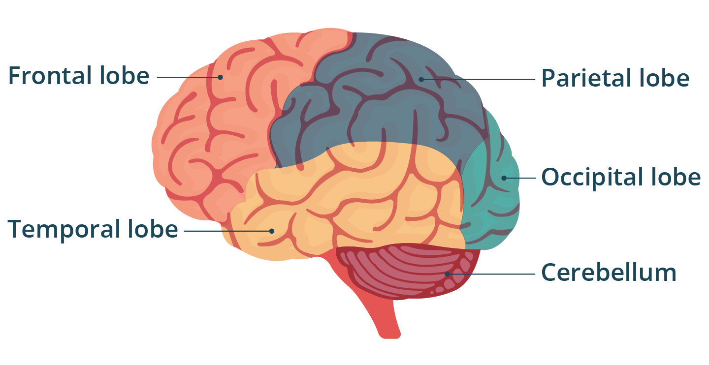
- Different regions (lobes) have specialized functions:
- Frontal lobe: decision-making, personality, voluntary motor control, speech production
- Parietal lobe: somatosensory processing, spatial awareness
- Temporal lobe: hearing, language comprehension, memory
- Occipital lobe: visual processing
Cerebellum:
- Located at the back of the brain, below the cerebrum
- Coordinates voluntary movements, balance, and posture
- Receives input from muscles, joints, and the inner ear
- Fine-tunes motor commands from the cerebrum, making movement smooth and precise
Brainstem:
- Connects the brain to the spinal cord
- Controls vital automatic functions:
- Medulla oblongata: breathing rate, heart rate, blood pressure, and swallowing
- Pons: relays signals between cerebrum and cerebellum, involved in breathing regulation
- Midbrain: visual and auditory reflexes, eye movement
Hypothalamus:
- Regulates homeostasis
- Controls the pituitary gland and therefore much of the endocrine system
- Regulates body temperature, hunger, thirst, sleep-wake cycles, and emotional responses
Thalamus: Acts as a relay station, routing sensory information from the body to the appropriate areas of the cerebral cortex
Limbic system:
- Involved in emotion, motivation, and memory formation
- Includes the hippocampus (memory) and amygdala (emotion, particularly fear)
Reflex Arcs
A reflex is a rapid, involuntary, stereotyped response to a stimulus that does not require conscious processing by the brain.
Reflexes are mediated by reflex arcs, neural pathways that bypass the brain, traveling only as far as the spinal cord before triggering a response.
The Spinal Reflex Arc Pathway
- The receptor detects the stimulus
- A sensory neuron carries an impulse to the spinal cord
- A relay neuron in the spinal cord connects sensory neurons to motor neurons (sometimes directly)
- Motor neuron carries an impulse to the effector
- The effector (muscle or gland) produces a response
Sensory information about the stimulus is also sent to the brain, but the reflex is complete before the brain responds.
Why Reflexes Bypass the Brain
The pathway through the spinal cord is far shorter than the path up to the brain and back. This dramatically reduces response time. For protective reflexes such as withdrawing from pain, this speed can prevent serious injury.
Types of Reflex
- Spinal reflexes: Mediated by the spinal cord. Examples include the knee-jerk reflex (patellar reflex) and the pain withdrawal reflex.
- Cranial reflexes: Mediated by the brainstem. Examples include the pupil light reflex and the gag reflex.
- Conditioned reflexes: Learned reflexes that develop through experience, famously demonstrated by Pavlov's dogs salivating at the sound of a bell associated with food.
Nervous System Disorders
Understanding the nervous system has clinical importance because many conditions involve its disruption.
- Multiple sclerosis: Autoimmune destruction of myelin sheaths, disrupting signal transmission. Causes progressive weakness, sensory disturbances, and coordination problems.
- Parkinson's disease: Progressive loss of dopamine-producing neurons in the midbrain, causing tremors, rigidity, and impaired movement.
- Epilepsy: Abnormal, synchronous electrical activity in groups of neurons causing seizures.
- Depression: Associated with disrupted neurotransmitter signaling, particularly serotonin and noradrenaline. Antidepressant medications target these neurotransmitter systems.
- Alzheimer's disease: Progressive loss of neurons, particularly in the hippocampus and cerebral cortex, causing memory loss, cognitive decline, and behavioral changes.