

Right now, somewhere in your body, a cell is dividing. Its chromosomes are being pulled apart by protein fibers. Its nuclear envelope has dissolved. Two new nuclei are forming. Within minutes, one cell will have become two.
This process happens billions of times in your body every day. It was built from a single fertilized egg. It heals your wounds. It replaces your worn-out blood cells. It is one of the most precisely choreographed processes in all of biology.
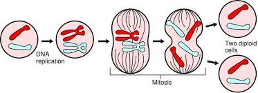
Mitosis is a type of cell division that produces two genetically identical daughter cells, each with the same number of chromosomes as the parent cell.
If the parent cell is diploid (2n), containing two sets of chromosomes, each daughter cell is also diploid (2n). In humans, where body cells contain 46 chromosomes, mitosis produces daughter cells each with 46 chromosomes, genetically identical to the parent.
Mitosis is used for growth, repair, and cell replacement in multicellular organisms, and for asexual reproduction in some organisms.
Before mitosis begins, the cell must copy all its DNA during the S phase of interphase.
After replication, each chromosome consists of two identical copies called chromatids, joined at a region called the centromere. These paired chromatids are called sister chromatids.
The cell now contains double the normal amount of DNA but still has the normal number of chromosomes, each consisting of two identical chromatids.
Prophase is the first and longest stage of mitosis.
Metaphase is the stage during which chromosomes are most clearly visible.
Metaphase is the stage at which chromosomes are most condensed and most clearly distinct, making it the best stage for counting chromosomes and studying their structure.
Anaphase is the stage during which chromosomes are separated.
Telophase is the final stage of mitosis.
Cytokinesis is the division of the cytoplasm, which usually occurs alongside or immediately after telophase.
In animal cells: A contractile ring of protein filaments pinches the cell membrane inward at the equator, eventually dividing the cytoplasm into two. This produces a cleavage furrow that deepens until the cell is separated into two daughter cells.
In plant cells: A cell plate forms across the middle of the cell from the inside outward. The cell plate is composed of vesicles from the Golgi apparatus that contain cell wall material. The cell plate expands until it fuses with the existing cell wall, dividing the cell into two.
| Stage | Key Events |
|---|---|
| Prophase | Chromosomes condense, nuclear envelope breaks down, and spindle forms |
| Metaphase | Chromosomes align at the cell equator, spindle fibers attach to centromeres |
| Anaphase | Sister chromatids separate and move to opposite poles |
| Telophase | Nuclear envelopes reform, chromosomes decondense |
| Cytokinesis | Cytoplasm divides, two daughter cells formed |
The two daughter cells produced by mitosis are:
This genetic identity is essential for mitosis's roles in growth and repair. When a skin cell divides to heal a wound, the new cells must be genetically identical skin cells, not some other cell type.
Some organisms reproduce entirely through mitosis, producing offspring that are genetically identical to the parent. This is called asexual reproduction.
Asexual reproduction through mitosis is rapid and efficient. However, all offspring are genetically identical, providing no genetic variation for natural selection to act upon.
Mitosis is carefully regulated. Cells normally divide only when signaled to do so by growth factors and only when conditions are appropriate.
Contact inhibition is the phenomenon by which cells stop dividing when they contact neighboring cells. This prevents overgrowth of tissues.
When the regulatory mechanisms controlling mitosis fail, cells may divide uncontrollably. This is the basis of cancer, where mutations in genes controlling the cell cycle lead to unregulated mitosis and tumor formation.