

Your heart will beat approximately 2.5 billion times during your lifetime. With each beat, it pushes blood through a network of vessels that, if laid end to end, would stretch approximately 100,000 kilometers. That is about two and a half times around the Earth.
This extraordinary circulatory system ensures that every one of your 37 trillion cells remains within a short diffusion distance of a blood supply, continuously receiving oxygen and nutrients and having waste products removed.
In very small organisms, diffusion is sufficient to move substances from the body surface to all internal cells. But as body size increases, the distances involved become too large for diffusion to be effective.
A molecule of oxygen diffusing through tissue takes approximately 1 second to move 100 micrometers. Crossing even a millimeter takes much longer. For an organism the size of a human, diffusion alone would take years to supply oxygen to internal cells.
The circulatory system solves this problem by rapidly transporting substances in bulk through a network of vessels, ensuring that no cell is more than a very short diffusion distance from a blood capillary.
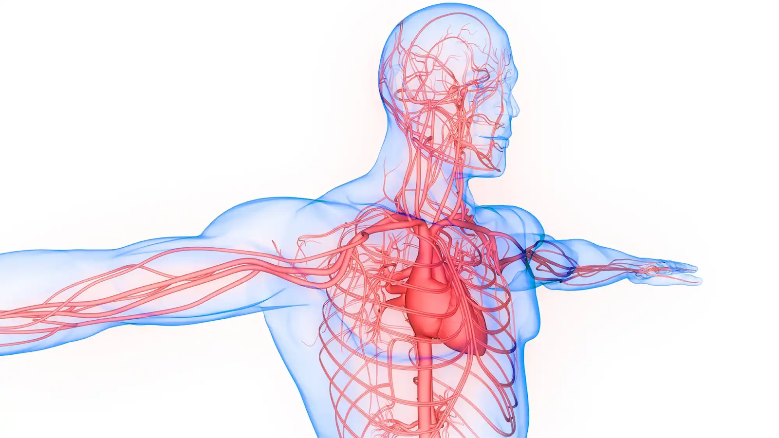
The human circulatory system consists of the heart, blood vessels, and blood. It is a closed, double circulatory system.
Closed means blood remains within vessels at all times and does not flow freely through body cavities.
Double circulation means blood passes through the heart twice for each complete circuit of the body. The right side of the heart pumps blood to the lungs (pulmonary circulation). The left side pumps blood to the rest of the body (systemic circulation).
Double circulation ensures that oxygenated blood reaches body tissues at high pressure, because it is repressurized by the left side of the heart after returning from the lungs. This allows efficient delivery of oxygen to all tissues.
The heart is a muscular pump located in the thoracic cavity between the lungs, slightly left of center.
The heart has four chambers.
Four valves ensure blood flows in one direction only through the heart.
Atrioventricular valves (bicuspid on the left, tricuspid on the right) separate the atria from the ventricles. They open when atria contract, allowing blood into ventricles, and close when ventricles contract, preventing backflow.
Semilunar valves at the exits of the ventricles (pulmonary and aortic valves) open when the ventricles contract, allowing blood out, and close when the ventricles relax, preventing backflow.
The sounds of the heartbeat ("lub-dub") are produced by these valves closing.
The cardiac cycle is the sequence of events during one complete heartbeat.
At rest, the cardiac cycle repeats approximately 70 times per minute. During exercise, it can increase to over 200 times per minute.
The heartbeat is initiated and coordinated by the heart's own electrical conduction system.
The rate of SA node firing, and therefore heart rate, is regulated by the autonomic nervous system and by hormones, particularly adrenaline, which increases heart rate during exercise and stress.
Three types of blood vessels form the vascular network.
Arteries carry blood away from the heart at high pressure.
Structure:
Function: The elastic walls stretch to accommodate the surge of blood with each heartbeat and recoil between beats, smoothing blood flow into a continuous stream rather than a series of pulses.
Arteries divide into smaller arterioles. The smooth muscle in arteriole walls controls their diameter, regulating blood flow to different organs.
Veins return blood to the heart at low pressure.
Structure:
Function: The large lumen reduces resistance to flow. Valves prevent backflow of blood, particularly important in the limbs where blood must flow upward against gravity. Blood is returned to the heart by the squeezing action of surrounding skeletal muscles during movement.
Capillaries are the smallest blood vessels and the sites where all exchange between blood and tissues occurs.
Structure:
Function: The one-cell-thick wall minimizes diffusion distance. The tiny diameter maximizes surface area relative to volume. The enormous total number ensures every cell is within approximately 100 micrometers of a capillary.
Oxygen, glucose, amino acids, hormones, and other substances diffuse out of capillaries into tissue fluid and then into cells. Carbon dioxide and metabolic waste products diffuse in the opposite direction from cells into capillaries.
Blood is a connective tissue consisting of cells and cell fragments suspended in a liquid matrix called plasma.
Plasma is pale yellow liquid constituting approximately 55 percent of blood volume.
Plasma transports:
Red blood cells are the most numerous blood cells, approximately 5 million per cubic millimeter.
Structural adaptations:
Function: Transport oxygen bound to hemoglobin. Each red blood cell contains approximately 280 million molecules of hemoglobin. Each hemoglobin molecule can carry four oxygen molecules. Hemoglobin loads oxygen in the high-oxygen environment of the lungs and releases it in the low-oxygen environment of active tissues.
White blood cells are fewer in number than red blood cells but far larger and more complex.
Phagocytes engulf and destroy pathogens, cellular debris, and foreign particles by phagocytosis.
Lymphocytes produce antibodies (B lymphocytes) or directly attack infected cells (T lymphocytes). They are the key cells of the specific immune response.
Platelets are small cell fragments derived from large bone marrow cells. They play a central role in blood clotting.
When a blood vessel is damaged, platelets adhere to the damaged site and aggregate, forming a platelet plug. They also release chemicals that initiate the clotting cascade, ultimately producing fibrin threads that reinforce the platelet plug and form a blood clot, preventing blood loss and providing a surface for tissue repair.
The cardiovascular system is vulnerable to disease, particularly atherosclerosis, the build-up of fatty deposits called plaques in arterial walls.
Plaques narrow the artery lumen, reducing blood flow. They can rupture, triggering clot formation that may completely block an artery. If this occurs in a coronary artery supplying the heart muscle, the result is a heart attack (myocardial infarction). If it occurs in an artery supplying the brain, the result is a stroke.
Risk factors for cardiovascular disease include high blood cholesterol, hypertension, smoking, physical inactivity, obesity, diabetes, and genetic predisposition.