

On this page:
Introduction Cell Theory Two Fundamental Types of Cells Animal Cell Structure Plant Cell Structure Animal Cell vs Plant Cell Microscopy and Magnification Structure Always Serves Function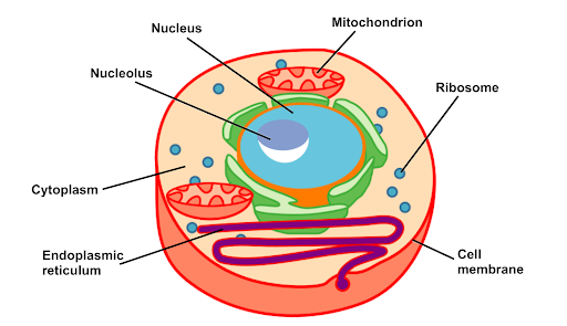
Have you ever wondered what you are actually made of? Not bones and blood in general, but the most basic unit of everything living. If you kept zooming in on your skin, your muscles, your brain, you would eventually reach something so small it is invisible to the naked eye. Something that is, despite its tiny size, one of the most complex and perfectly organized structures in the known universe.
That something is a cell.
A cell is the basic structural and functional unit of all living organisms. Every living thing on Earth, from the smallest bacterium to the largest blue whale, is made of cells. Some organisms consist of just one single cell doing everything needed to survive. Others, like humans, are built from approximately 37 trillion cells, each with a specific job, all working together in extraordinary coordination.
Understanding cell structure and function is not just one biology topic among many. It is the foundation on which all of biology is built.
Before exploring the structure of cells, it is important to understand the three principles that form the foundation of all cell biology. These are collectively called the cell theory.
These principles were established in the 19th century through the work of Matthias Schleiden, Theodor Schwann, and Rudolf Virchow. They remain among the most firmly supported ideas in all of science.
Not all cells are built the same way. Every cell on Earth belongs to one of two broad categories based on whether they have a membrane-bound nucleus.
A prokaryotic cell is a cell that does not have a membrane-bound nucleus. Its genetic material, DNA, floats freely in the cytoplasm in a region called the nucleoid. Prokaryotes are always unicellular. Bacteria are the most well-known example. These cells are generally small, typically between 1 and 10 micrometers in diameter.
A eukaryotic cell is a cell that contains a membrane-bound nucleus housing its DNA. Eukaryotic cells are larger and more structurally complex than prokaryotic cells. All multicellular organisms, including plants, animals, and fungi, are made of eukaryotic cells.
An animal cell contains a number of specialized structures called organelles. The word organelle means "little organ," and just like organs in the body, each organelle has a specific structure and a specific function. Understanding both together is the key to cell biology.
Structure: The cell membrane is a thin, flexible layer completely surrounds every cell. It is made of a double layer of phospholipid molecules called the phospholipid bilayer, with protein molecules embedded within and across it.
Function: The cell membrane controls what enters and leaves the cell. It is selectively permeable, meaning it allows some substances to pass through freely while blocking others.
This selective control is critical for maintaining the correct internal environment the cell needs to function.
Structure: The nucleus is a large, roughly spherical organelle surrounded by a double membrane called the nuclear envelope. The envelope contains small openings called nuclear pores. Inside, DNA is organized into structures called chromosomes. A dense region within the nucleus called the nucleolus is also present.
Function: The nucleus is the control center of the cell. It contains the genetic instructions in the form of DNA for building every protein the cell produces.
Structure: The cytoplasm is the gel-like fluid called cytosol that fills the interior of the cell and surrounds all the organelles.
Function: Provides the medium in which organelles are suspended; allows substances to move around the cell; site of many chemical reactions including glycolysis, the first stage of cellular respiration.
Structure: Mitochondria are oval-shaped organelles surrounded by two membranes. The inner membrane is folded inward into structures called cristae. The space enclosed by the inner membrane is called the matrix.
Function: Mitochondria carry out aerobic cellular respiration, producing ATP, the molecule that provides energy for all cellular activities.
Structure: Ribosomes are extremely small organelles made of ribosomal RNA and proteins. They are found either floating freely in the cytoplasm or attached to the surface of the endoplasmic reticulum.
Function: Ribosomes carry out protein synthesis, building proteins by reading instructions carried by mRNA from the nucleus.
Structure: The endoplasmic reticulum is an extensive network of folded membranes extending from the nuclear envelope throughout the cytoplasm. Rough ER has ribosomes on its outer surface. Smooth ER has no ribosomes.
Function:
Structure: The Golgi apparatus consists of a stack of flattened, curved membrane-bound sacs called cisternae.
Function: The Golgi apparatus is the cell's packaging, sorting, and distribution center.
Structure: Lysosomes are small, spherical organelles surrounded by a single membrane, produced by the Golgi apparatus. They maintain an acidic internal environment.
Function: Lysosomes are the cell's waste disposal and recycling system.
Structure: Centrioles are small, cylindrical structures found in pairs near the nucleus in animal cells, forming a structure called the centrosome.
Function: Organize spindle fibers during cell division; spindle fibers attach to chromosomes and pull them apart to opposite ends of the cell; ensure each daughter cell receives the correct number of chromosomes. Found in animal cells but absent from most plant cells.
Plant cells share all the organelles found in animal cells. However, they have three additional structures that are essential to their unique way of life.
Structure: The cell wall is a rigid outer layer surrounding the cell membrane of every plant cell. It is composed primarily of cellulose, a complex carbohydrate made of long chains of glucose molecules arranged in strong fibers.
Function: Provides structural support and mechanical strength to the plant cell; allows plants to grow tall without a skeleton; prevents the cell from bursting when water enters by osmosis; creates turgor pressure that keeps plant tissues firm and upright. Fully permeable and does not restrict what passes through it.
Structure: Chloroplasts are large, oval-shaped organelles surrounded by a double outer membrane. Inside, flattened sac-like structures called thylakoids are stacked in columns called grana. The fluid surrounding the grana is called the stroma.
Function: Chloroplasts are the sites of photosynthesis.
Structure: The central vacuole is a large, membrane-bound sac filled with cell sap, a solution of water, sugars, salts, and other dissolved substances. In a mature plant cell it can occupy up to 90 percent of the cell's total volume.
Function: Maintains turgor pressure by filling with water and pushing against the cell wall; keeps plant tissues firm and upright; stores nutrients, waste products, and pigments; in some plants, there are toxic compounds that deter animals from eating the plant.
| Feature | Animal Cell | Plant Cell |
|---|---|---|
| Cell membrane | Present | Present |
| Nucleus | Present | Present |
| Mitochondria | Present | Present |
| Ribosomes | Present | Present |
| Cell wall | Absent | Present (cellulose) |
| Chloroplasts | Absent | Present |
| Central vacuole | Small or absent | Large and permanent |
| Lysosomes | Present | Rarely present |
| Centrioles | Present | Absent in most |
Cells are far too small to see with the naked eye. Their study depends entirely on microscopes.
Magnification is calculated as: Magnification = Image size divided by Actual size
In cell biology, nothing exists by accident. Every structural feature of every organelle is directly connected to its function. The folded cristae of mitochondria maximize surface area for energy production. The ribosomes on the rough ER position protein production directly inside the transport system. The acidic interior of lysosomes provides the optimal pH for their digestive enzymes.
When you understand both structure and function together, the cell stops being a list of parts to memorize. It becomes a remarkably logical, elegantly organized system that makes complete sense.