

On this page:
Introduction Nervous System Structure of Neuron Types of Neurons Myelination and Saltatory Conduction Resting Membrane Potential Action Potential Action Potential Stages Action Potential Propagation Synapse and Synaptic Transmission Neurotransmitters Summation - Integration of multiple signals Neuromuscular Junction Neural Signaling in Reflexes Disorders Related to Neural Signaling Energy and Neural Signaling Significance of Neural Signaling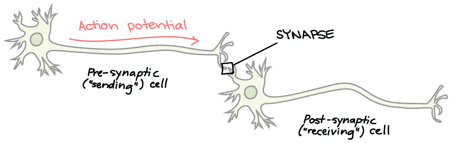
Example: touch pan, hot pain, pull hand away, system reaction, brain identification of pain
As one of the most important systems in the human body, the speed of the communication system is lightning fast. Whereas with the pace of the world, it is as fast as the speed of light, communication systems are made in the world. In the body, instead of wires, there are neurons that transport the messages. In Biology, Neural Signaling is the critical answer to the question of how the body responds to stimuli and maintains balance.
Think of the Nervous system as a communication system in the body. It is a system that responds and controls the stimuli in the environment. The system is made of 2 Main parts:
The motor division may be classified as follows:
Neurons are the most basic building blocks of the nervous system. Neurons are specifically structured to carry signals from one cell to another. Typical neurons are made up of 3 components:
There are three classes of neurons based on specific functions.
Several axons are encased within a fatty layer known as the myelin sheath. In the CNS, myelin is produced by oligodendrocytes, while in the PNS, myelin is produced by Schwann cells.
The resting membrane potential is the first stage of neural signaling. The septum of a neuron separates two different environments: inside the cell and outside of the cell. The neuron will be resting, and there will be a net negative charge inside the neuron as opposed to the outside - this is called the resting membrane potential and is typically about -70 mV.
The reason for this difference in charge is due to something called the ion imbalance. This phenomenon is explained with respect to three things: The active transport of the different cell membrane components, as well as the sodium–potassium pump.
The sodium–potassium pump transports 3 Na⁺ ions out of the cell while bringing 2 K⁺ ions into it, utilizing ATP energy from the cell.
This increases the concentration gradient in the cell as well as the net negative charge in the cell.
An action potential occurs when there is a significant increase in the charge (positive and negative) across the membrane, which is a phenomenon that occurs as the impulse travels along the axon. This is called an all-or-none phenomenon, which means that in response to a stimulus, there will be an action potential, irrespective of the stimulus.
If a stimulus does not reach a certain voltage threshold, action potentials will not be generated.
The threshold is typically around -55 mV. When this threshold is reached, an action potential is triggered.
The action potential will cascade down the axon, as the depolarization of one segment will cause the depolarization of the next segment.
Neurons do not directly touch, and the gap between them is called a synapse.
A synapse has three components:
Synaptic transmission is a chemical process. When an action potential gets to the end of an axon:
Neurotransmitters are molecules that transmit messages. Some examples are:
When a neurotransmitter attaches to a receptor, its effect may be:
Following transmission, neurotransmitters:
A single EPSP may not reach the threshold. However, a neuron integrates multiple signals to reach a threshold.
Summation comes in two forms:
The moment the total depolarization hits the threshold, an action potential will be born.
A unique synapse that connects a motor neuron with a muscle fiber is called a neuromuscular junction.
When acetylcholine is released:
When a muscle contracts, the process involves a step called depolarization, the synapse that causes the contraction releases a chemical messenger known as acetylcholine, and to stop the signal, an enzyme called acetylcholinesterase comes into play.
Reflexes occur without conscious thought.
The reflex arc involves:
Reflexes are an important part of bodily protection.
Proper neural signaling ensures that the body functions normally.
Neurons need constant amounts of oxygen supplied by the blood, as well as glucose. Energy is also needed in the process of ATP formation.
This is required in:
When the energy is absent, the signaling process ceases to exist.
Neurons change the body's original system to adapt to the new environmental changes. The body works as a system, and neural signaling is the most important part of the system that ensures secure, precise, coordinated movement and control of all the internal factors.
The nervous system helps in planning, organizing, and remembering activities. Neural signaling can turn the electrical energy in the body into a chemical energy signal. The neural system works by exerting concentration, or energy of the body.
Neural signaling from resting membrane potential to action potential, from synaptic transmission to reflex action, describes the system of millions of neurons functioning in unison to bring about behavior, sensitivity, and consciousness.