Muscles and Motility
Every time you run, lift a bag, or even blink your eyes, muscles are helping you do these activities. Muscles are a type of tissue in your body. They help generate movement and are critical even for activities you may not consciously control, like your heart beating, or food and your muscles move through your intestines.
In this lesson, we will examine the different types of muscles and their structures and also how motility occurs at the cellular level and also at the level of the entire body.
Types of Muscles
Three muscle types exist in the human body:
Skeletal Muscle
- Skeletal muscles are attached to bones using a tendon
- There are voluntary movements that can be controlled
- There is a striated appearance due to the organisation of the protein filaments
- The cells are multinucleated for efficient production and repair of proteins
Cardiac Muscle
- This muscle is found in the heart only
- It is involuntarily controlled and works without conscious control
- Striated cells that are branched and have intercalated discs that facilitate uniform contraction
Smooth Muscle
- These are located in organs like the intestine, blood vessels, and the bladder
- The muscle movements are involuntary
- Their structure has no striations and has cells that resemble a spindle
- These muscles may contract slowly, but they are able to maintain the contraction for long durations
Structure of Skeletal Muscle
The tissue is made of many specialized units, and the units are organized in a particular manner and can be described as follows:
- Muscle Fiber (Cell): Individual units that are long and cylindrical and have several nuclei.
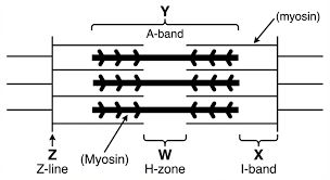
- Myofibrils: These are bundles and are found in muscle fibers and have structures called sarcomeres that repeat in units.
- Sarcomere: These are the units responsible for contraction and have two types of filaments: actin (thin) and myosin (thick) filaments.
When you examine the cells in a muscle, the many sarcomeres in a row give the tissue a striated look.
The Sliding Filament Theory
So, how does a muscle achieve contraction? The answer is found in the sliding filament theory:
- Muscle contraction involves the sliding action of actin and myosin past each other.
- The myosin heads that have bound ATP can cross from one actin to the other and 'walk' towards the center of the sarcomere.
- As a result, the length of the sarcomere is reduced, and contraction is achieved without any change to the length of the filaments.
Main Points:
- ATP interacts with myosin to enable the pulling and releasing of actin.
- Calcium ions (Ca²⁺) regulate the contraction process by covering actin with binding sites.
Neuromuscular Junction
A skeletal muscle contracts only when it is stimulated by the nervous system.
- A motor neuron is in direct contact with a muscle fiber at the NMJ.
- An action potential on the neuron leads to the exocytosis of the neurotransmitter acetylcholine (ACh) into the synaptic cleft.
- ACh binds to receptors on the sarcolemma (the muscle cell membrane) and initiates depolarization.
- The depolarization is transmitted through the T-tubules that cause the release of Ca²⁺ from the sarcoplasmic reticulum.
- Calcium attaches to troponin, thereby making actin available for myosin binding, and initiates contraction.
Muscle Contraction Cycle
The contraction cycle has the following steps:
- Resting state: Myosin heads are charged, and the binding sites for actin are temporarily inactive with tropomyosin.
- Cross-bridge formation: Calcium ions become available, and binding sites are unblocked; myosin binds to actin.
- Power stroke: Myosin pulls the actin, which causes the sarcomere to shorten. It results in the release of ADP.
- Detachment: Muscle contraction is stopped when ATP attaches to myosin, and myosin is released from actin.
- Reactivation: ATP is used, and the myosin cross-bridge is reset to the energized state.
When calcium (Ca²⁺) is reabsorbed into the sarcoplasmic reticulum, actin binding sites are again covered, and relaxation is over.
Types of Skeletal Muscle Fibers
Slow-twitch (Type I)
- Contain more mitochondria and myoglobin and are red
- Provide high endurance but low contraction speed
- Example: Long-distance running
Fast-twitch (Type II)
- Fewer mitochondria, more pale in appearance
- Contract quickly and powerfully
- Example: Sprinting, weightlifting
Energy for Muscle Contraction
Muscles require ATP, which powers myosin. ATP comes from:
- Creatine phosphate: Like the refresh button, but lasts for only a few seconds.
- Anaerobic respiration: Gives lactic acid as a byproduct.
- Aerobic respiration: Occurs in the mitochondria for sustained energy production.
Motility at the Cellular Level
Motility goes beyond just the muscles. Cells also move using specialized structures:
Flagella
Longer whip-like structures used for propulsion in single-celled organisms and sperm cells.
Cilia
Short, hair-like structures that move in coordinated waves, like oars, to move fluids or the cell itself.
Amoeboid Movement
Cells extend pseudopodia (false feet) to crawl along surfaces. This type of movement does not rely on myosin in the same way as skeletal muscle contraction.
Muscle Coordination and Movement
The body cooperates through mutual antagonism in pairs:
- Muscles only pull; they cannot push.
- One muscle constricts while the other relaxes to create movement.
Example:
- Biceps: Arm flexes (contracts)
- Triceps: Arm extends (relaxes)
Disorders Affecting Muscles
Understanding muscles also involves what can go wrong:
Muscular Dystrophy
A defective protein leads to muscular dystrophy, a genetic disease that weakens the muscles progressively over time.
Tetanus
An uncontrolled contraction caused by the toxin of a bacterium, leading to painful muscle spasms.
Myasthenia Gravis
An autoimmune disorder that leads to weakness due to the blockage of the acetylcholine (ACh) receptors at the neuromuscular junction.
Summary
- Muscles are specialized tissues that are designed for motion.
- There are three types of muscles: skeletal (voluntary), cardiac (heart), and smooth (organs).
- Contraction of skeletal muscles is based on the sliding filament theory.
- Contraction is initiated by the neuromuscular junctions and Ca²⁺ (calcium) ions.
- Some muscle fibers are designed for endurance, while others are designed for rapid movement.
- ATP can be supplied through a variety of sources: creatine phosphate, anaerobic pathways, and aerobic pathways.
- At the organismal and cellular levels, structures such as cilia, flagella, and pseudopodia are used for movement.
- The coordination of the muscles demonstrates the importance of the proper functioning of muscles and the disorders that impede motion.
Understanding muscles and motility demonstrates the movement of life, from the microscopic movement of the cells to the movement of the entire body. Muscle is tissue, but it is also the tissue of life.